45 ear diagram no labels
20 Best Types of Ear Piercings: Styles, Pain Chart & Costs ... 2 Different Types of Ear Piercings 2.1 Industrial Piercing 2.2 Ear Lobe Piercing 2.3 Cartilage Piercing 2.4 Helix Piercing 2.5 Forward Helix Piercing 2.6 Daith Piercing 2.7 Tragus Piercing 2.8 Snug Piercing 2.9 Transverse Lobe Piercing 2.10 Orbital Piercing 2.11 Anti-Tragus Piercing 2.12 Conch Piercing 2.13 Rook Piercing 2.14 Top Ear Piercing Eye Diagram Quiz - ProProfs Settings Create your own Quiz Questions and Answers 1. What is 1? A. Ciliary body B. Cornea C. Iris D. Aqueous humor 2. What is 2? A. Sclera B. Retina C. Suspensory ligaments D. Optic nerve 3. What is 3? A. Iris B. Lens C. Iridociltis D. Sclera 4. What is 4? A. Iris B. Cornea C. Lens D. Pupil 5. What is 5? A. Anterior chamber B. Pupil C. Lens D.
Cow Anatomy - External Body Parts and Internal Organs with ... The external body parts from the head region of a cow - in this head region, you might identify the mouth, lip, cheek, chin, muzzle, forehead, poll, ear, eye, nostril, and other. Different parts from the neck region of a cow - here, you will find the neck crest, dewlap, brisket, and jugular groove.

Ear diagram no labels
What are the different parts and functions of an otoscope? References. Answer. An otoscope consists of a head and a handle and is used to examine the external auditory canal (EAC), the tympanic membrane, and the middle ear. A magnifying lens enhances the ... Parts and Components of Human Ear and Their Functions | MD ... In addition to helping the body take in auditory messages, the ear helps to maintain a proper head position. The fluid in the ear also helps the body maintain a sense of balance so the body can maintain proper posture and coordination. There are three major parts of the ear, the outer, middle and inner ear. Each contains several parts that are ... › pin › 391461392603795120Image result for ear structure without label | Ear diagram ... Feb 12, 2018 · Parts of the Eye Diagram for 4th graders | Lesson 2 Grade 3 - Grade 4 Activities. The olfactory system enables us to detect odors. Our sense of smell involves nerves, the brain, and sensory organs such as the nose and olfactory bulbs. Original Dust Brush is a good, comfortable, fast - to - use product.
Ear diagram no labels. Structure and Functions of Human Eye with labelled Diagram The External Structure of an Eye. Sclera: It is a white visible portion. It is made up of dense connective tissue and protects the inner parts. Conjunctiva: It lines the sclera and is made up of stratified squamous epithelium. It keeps our eyes moist and clear and provides lubrication by secreting mucus and tears. Labeled atlas of anatomy: illustrations of the dog - IMAIOS Anatomy of the dog - Illustrated atlas. This modules of vet-Anatomy provides a basic foundation in animal anatomy for students of veterinary medicine. This veterinary anatomical atlas includes selected labeling structures to help student to understand and discover animal anatomy (skeleton, bones, muscles, joints, viscera, respiratory system ... The ear canal: Anatomy, diagram, and common conditions Conductive hearing loss is a form of hearing impairment that involves the outer ear. Any type of obstruction in the ear canal, including stenosis, growths, and infections, can be a potential cause... Examining and medicating a cat's ears | Veterinary ... How to hold your cat while cleaning or medicating its ears. To hold your cat in your lap to place ear medications, drape your left forearm across the cat's body to keep it in your lap. Hold the head with your left hand using your left thumb to press the ear flap against the head with the ear canal open. Hold the medication in your right hand.
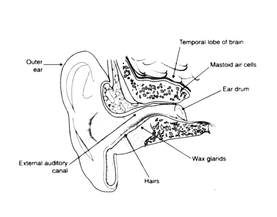
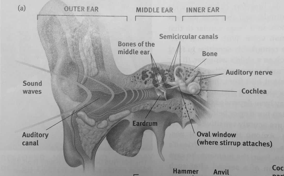
ER Diagram: Entity Relationship Diagram Model | DBMS Example ER Model stands for Entity Relationship Model is a high-level conceptual data model diagram. ER model helps to systematically analyze data requirements to produce a well-designed database. The ER Model represents real-world entities and the relationships between them. Creating an ER Model in DBMS is considered as a best practice before ... Examining and medicating the ears of a dog | Veterinary ... The pictures above show a diagram of the right ear as it appears if you are looking at the dog's head from the front and a CT scan of the head. The outer ear flap is usually covered with fur. If the ear is itchy, scratching may result in hair loss on the ear flap or at the base of the ear. Severe scratching may also lead to tears at the edges ... Ear diagram labelling game - ESL Games Plus The ear itself is an intricate organ divided into three primary parts - the outer ear, middle ear, and inner ear. Sound is first collected by the pinna - this is the visible part of the ear we typically see on our heads. The pinna gathers and funnels the sound into the ear canal, causing it to strike the eardrum. Diagram of Human Heart and Blood Circulation in It | New ... Every heart diagram labeledwill clearly show these valves. These valves allow blood flow in one direction only. Different valves perform different functions. Tricuspid valve is located between the right ventricle of your heart and the right atrium, and allows the blood to move from the right atrium to the right ventricle.
› en › libraryBlank ear diagrams and quizzes: The fastest way to learn - Kenhub Oct 28, 2021 · Take a moment to look at the ear model labeled above. This shows you all of the structures you’ve just learned about in the video, labeled on one diagram. Seeing them all together in this way is a great way to learn, since anatomical structures do not exist in isolation. That’s why labeling the ear is an effective way to begin your revision. Ear Diagram Quiz - ProProfs Ear Diagram Quiz. 14 Questions | By Bellamiller123 | Last updated: Mar 22, 2022 | Total Attempts: 5610. Questions All questions 5 questions 6 questions 7 questions 8 questions 9 questions 10 questions 11 questions 12 questions 13 questions 14 questions. Settings. Feedback. During the Quiz End of Quiz. Difficulty. Sequential Easy First Hard First. Anatomy, Head and Neck, Ear Eustachian Tube - NCBI Bookshelf Patency of the tube allows for air exchange in the tympanic cavity to replenish oxygen to the middle ear, in addition to providing an outlet for mucus and other fluid from the middle ear. [3] [4] Partly a hollow tube in bone and partly a potential space in fibroelastic cartilage, the Eustachian tube is normally closed, as its proximal walls are ... Ear Anatomy Illustration - human ear anatomy stock vector ... Ear Anatomy Illustration - 16 images - hair cells in a mammal cochlea the portion of the inner, ear anatomy stock image c022 1021 science photo library, ear anatomy the ear is the organ that detects sound it, ear anatomy clip art k40993746 fotosearch,
anatomy, ear diagram to label | Middle School General Music | Pinterest | Words, Science and ...
Ileum Histology Slide with Labeled Diagram and ... The wall of the ileum histology consists of four distinguished layers - tunica mucosa, submucosa, muscular, and serosa. You will find thin and slender villi on the tunica mucosa layer of the ileum slide. The simple columnar epithelium lines these villi of the ileum. So, you will find the intervillous spaces between the villi of the ileum structure.

Human ear Royalty Free Vector Image - VectorStock
Trigeminal Nerve: Function, Anatomy, and Diagram Function. The trigeminal nerve is the largest of the 12 cranial nerves. Its main function is transmitting sensory information to the skin, sinuses, and mucous membranes in the face. The nerve ...
35 Diagram Of The Ear To Label - Labels Design Ideas 2020
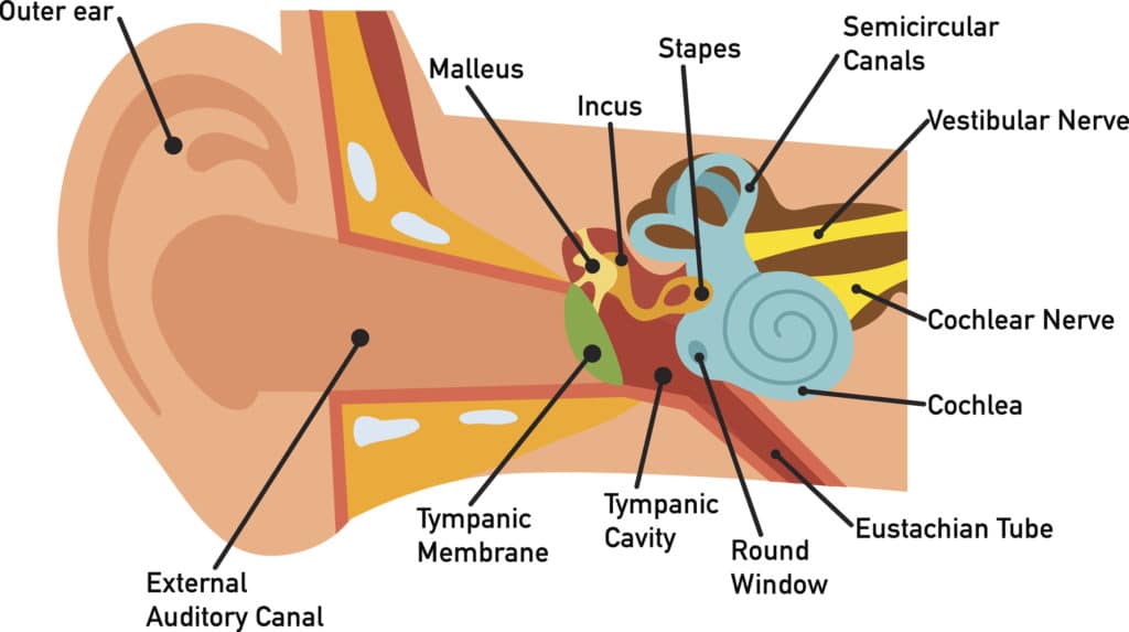
Malleus: Anatomy, Function, and Treatment - Verywell Health Anatomy. The malleus ("hammer"), incus ("anvil"), and stapes ("stirrup") are the three bones, also known as ossicles, of the inner ear. The malleus is the largest and the outermost of the bones, which are part of the auditory system. Together, the three bones make up an area no larger than the seed of an orange.

33 Label Ear Diagram - Labels For Your Ideas
Diagram Of The Heart Not Labeled Ear Diagram No Lines Wiring Diagram Schematic Name . Human Body Diagram Not Labeled Modern Design Of Wiring Diagram . Diagram Of Plant Cell With Labels Simple Animal For Kids Not Labeled . External Heart Diagram Not Labeled . 19 Heart Diagram Templates Sample Example Format Download ...

How You Hear - Northland Audiology
Parts of Stethoscope: A Comprehensive Overview 1-Headset. The headset is a part of a stethoscope, which is a combination of ear tips and ear tubes, and tension springs. These components combine together to fulfilling the purpose of diagnosis. It provides a comfortable alignment in the ears of a user and further used to provide maximum quality of sound through the headset.
How You Hear - Northland Audiology
conchahearing.com › interactive-ear-diagramEar Diagram - Concha Audiology The cochlea is a fluid-filled organ essential for the transduction of mechanical (vibration) energy to electrical (nerve impulse) energy. Vibrations from the stapes on the oval window cause waves within the fluid, which causes the basilar membrane to move. The movement of the basilar membrane causes a shearing action of hair cells (outer and ...
31 Label The Ear Anatomy Diagram Answers - Labels For You
Human Ear Diagram Blank - a p, ear diagram video human ... Human Ear Diagram Blank. Here are a number of highest rated Human Ear Diagram Blank pictures on internet. We identified it from trustworthy source. Its submitted by admin in the best field. We understand this nice of Human Ear Diagram Blank graphic could possibly be the most trending subject behind we part it in google plus or facebook.
Ear Diagram With Labels | PowerPoint Shapes | PowerPoint Slide Deck Template | Presentation ...
Labelled imaging anatomy cases | Radiology Reference ... URL of Article. This article lists a series of labelled imaging anatomy cases by body region and modality. On this page: Article: Brain. Head and neck. Spine. Chest. Abdomen and pelvis.

Our TN Adventure: Disbudding & Tattooing the Goats
byjus.com › biology › diagram-of-earWell-Labelled Diagram Of Ear With Explanation - BYJUS Well-Labelled Diagram of Ear. The External ear or the outer ear consists of: Pinna/auricle is the outermost section of the ear. The external auditory canal links the exterior ear to the inner or the middle ear. The tympanic membrane, also known as the eardrum, separates the outer ear from the inner ear. The Middle ear comprises:

Label Ear Diagram - Diagram Resource Gallery
Sense Organs ICSE Class 10 Biology Board Exam Questions The tube which connects the cavity of the middle ear with the throat Ans. 14. Transmits impulse to the brain from the ear Ans. 15. Part of your hand which is most sensitive to touch Ans. 16. The photoreceptors found in the retina of the eyes Ans. 17. The eye defect caused due to shortening of the eye ball from front to back Ans. 18.
Hansen Psychology - Just Keeping Things Here
Anatomical Line Drawings - Medscape Surface Anatomy - lateral views - male. go to drawing without labels. Surface Anatomy - lateral views - female. go to drawing without labels. Surface Anatomy - Child - anterior view & posterior ...
Ear | ClipArt ETC
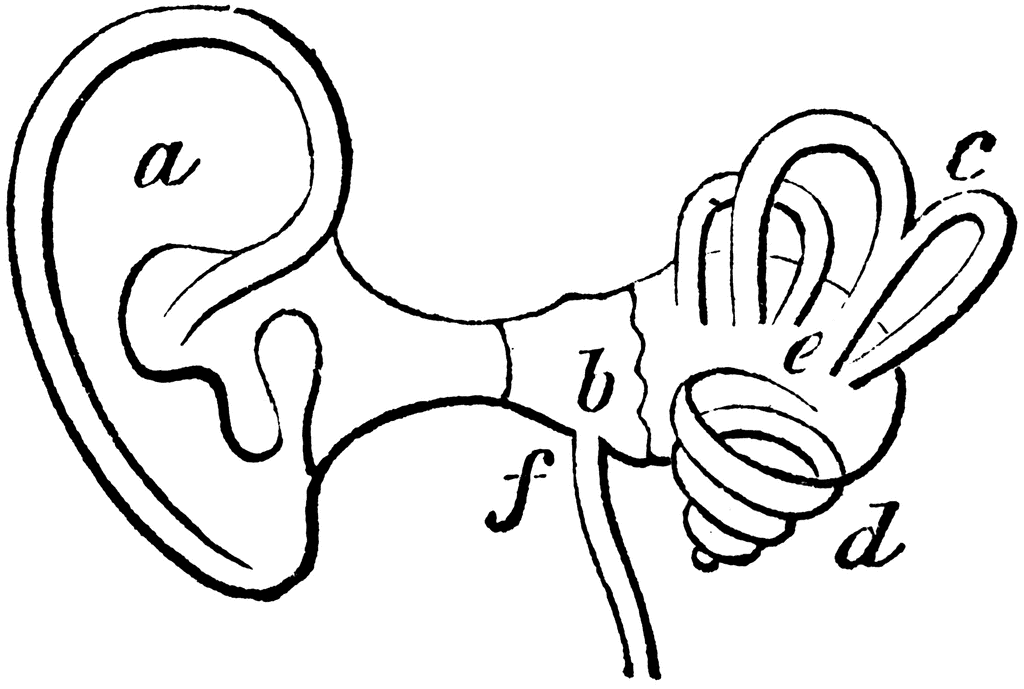
Ear anatomy: Parts and functions - Kenhub The ear is anatomically divided into three portions: External ear Middle ear Internal ear This mixture of bones, nerves, vessels, membranes, and muscles that make up the ear will be described in this article. Contents External ear Auricle External acoustic meatus Tympanic membrane Muscles of the external ear Vasculature of the external ear

Pin by elizabeth wright on Free time | Human skull anatomy, Skull anatomy, Skull
Eustachian Tube: Anatomy, Location, and Function The eustachian tube consists of bone, cartilage, and fibrous tissue. The hollow tube is lined with cilia, hair-like projections that sweep mucus away from the middle ear toward the nasopharynx. 1. Six muscles contribute to the opening and closing of the eustachian tube. They are located in the ear, head, neck, soft palate, and jaw. 1.
Ear - Teaching resources
› pin › 391461392603795120Image result for ear structure without label | Ear diagram ... Feb 12, 2018 · Parts of the Eye Diagram for 4th graders | Lesson 2 Grade 3 - Grade 4 Activities. The olfactory system enables us to detect odors. Our sense of smell involves nerves, the brain, and sensory organs such as the nose and olfactory bulbs. Original Dust Brush is a good, comfortable, fast - to - use product.

Biology Diagrams,Images,Pictures of Human anatomy and physiology: Complete ear structure
Parts and Components of Human Ear and Their Functions | MD ... In addition to helping the body take in auditory messages, the ear helps to maintain a proper head position. The fluid in the ear also helps the body maintain a sense of balance so the body can maintain proper posture and coordination. There are three major parts of the ear, the outer, middle and inner ear. Each contains several parts that are ...

Human Anatomy Lab: Ear Models
What are the different parts and functions of an otoscope? References. Answer. An otoscope consists of a head and a handle and is used to examine the external auditory canal (EAC), the tympanic membrane, and the middle ear. A magnifying lens enhances the ...

Hearing Loss - Dallas ENT Group
Post a Comment for "45 ear diagram no labels"