39 microscope images with labels
Label the microscope — Science Learning Hub All microscopes share features in common. In this interactive, you can label the different parts of a microscope. Use this with the Microscope parts activity to help students identify and label the main parts of a microscope and then describe their functions. Drag and drop the text labels onto the microscope diagram. Microscope Labeling - The Biology Corner The google slides shown below have the same microscope image with the labels for students to copy. I often spend the first day walking students through the steps and having them look at a single slide as we do the steps. Students are often very enthusiastic about using microscopes and will try to start with the high power objective.
› products › microscopeLAS X Industry Microscope software for Industry | Products ... Create a single sharp image by capturing a stack of images at different focus positions and combining them automatically into an Extended Depth of Focus (EDOF) image. LAS X Extended Depth of Field: Create sharp 2D images from several partially in-focus images. In connection with the 3D Surface Viewer, creation of 3D images is also possible.

Microscope images with labels
Compound Microscope Parts, Functions, and Labeled Diagram Compound Microscope Definitions for Labels. Eyepiece (ocular lens) with or without Pointer: The part that is looked through at the top of the compound microscope. Eyepieces typically have a magnification between 5x & 30x. Monocular or Binocular Head: Structural support that holds & connects the eyepieces to the objective lenses. Polarizing Microscope Image Gallery | Science Lab - Leica Microsystems Polarized light microscopy (also known as polarizing microscopy) is an important method used in different fields, including research and quality assurance. It goes beyond just producing images at high magnification and resolution, something typically done with microscopes using ordinary optics. By examining the form, structure, color, birefringence, and further optical properties, additional ... › WAI › EMIndex of Dr.Jastrow's electron microscopic atlas Table D leads to images of electron microscopes or protocols for tissue preparation. Table E leads to the overview pages with the images of this atlas which are used in the histology course of the University of Mainz, Germany. From table F you can call up the Vocabulary of microscopic anatomy which explains some terms in German and Englisch.
Microscope images with labels. › Microscope-ANNLOV-ElectronicAmazon.com : LCD Digital Microscope,ANNLOV 4.3 inch Handheld ... Feb 05, 2020 · This item LCD Digital Microscope,ANNLOV 4.3 inch Handheld USB Microscope 50X-1000X Magnification Coin Microscope Video Camera with 8 Adjustable LED Lights for Adults PCB Soldering Kids Outside Use ANNLOV 7" LCD Digital Microscope with 32GB TF Card 1200X Maginfication 1080P Coin Microscope with Wired Remote,12MP Ultra-Precise Focusing Video ... microscope picture with labels - Compound Light Microscope... View microscope picture with labels from BIOL 1005Y at Yeshiva University. Compound Light Microscope ocular (eyepiece) revolving nosepiece objectives coarse adjustment knob mechanical stage fine 400+ Free Microscope & Bacteria Images - Pixabay 412 Free images of Microscope Related Images: bacteria laboratory science scientist research biology lab virus microscopic Find your perfect microscope image. Free pictures to download and use in your next project. Parts of a microscope with functions and labeled diagram - Microbe Notes Optical parts of a microscope and their functions The optical parts of the microscope are used to view, magnify, and produce an image from a specimen placed on a slide. These parts include: Eyepiece - also known as the ocular. This is the part used to look through the microscope. Its found at the top of the microscope.
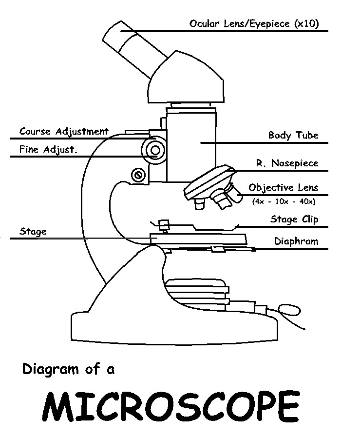
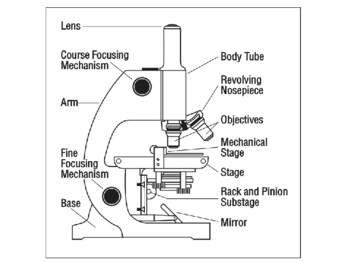
Microscope, Microscope Parts, Labeled Diagram, and Functions Revolving Nosepiece or Turret: Turret is the part of the microscope that holds two or multiple objective lenses and helps to rotate objective lenses and also helps to easily change power. Objective Lenses: Three are 3 or 4 objective lenses on a microscope. The objective lenses almost always consist of 4x, 10x, 40x and 100x powers. The most common eyepiece lens is 10x and when it coupled with ... Microscope With Labels clip art | Microscope parts, Scientific method ... clker.com vector clip art online, royalty free & public domain Download Clker's Microscope With Labels clip art and related images now. Multiple sizes and related images are all free on Clker.com. D Dixie Tsutsaeva 2k followers More information Microscope With Labels clip art Find this Pin and more on Art Journal Inspiration by Dixie Tsutsaeva. Parts of the Microscope with Labeling (also Free Printouts) Microscopes are specially created to magnify the image of the subject being studied. This exercise is created to be used in homes and schools. the microscope layout, including the blank and answered versions are available as pdf downloads. Click to Download : Label the Parts of the Microscope (A4) PDF print version. A Study of the Microscope and its Functions With a Labeled Diagram A Study of the Microscope and its Functions With a Labeled Diagram To better understand the structure and function of a microscope, we need to take a look at the labeled microscope diagrams of the compound and electron microscope. These diagrams clearly explain the functioning of the microscopes along with their respective parts.
rsscience.com › stereo-microscopeParts of Stereo Microscope (Dissecting microscope) – labeled ... Unlike a compound microscope that offers a flat image, stereo microscopes give the viewer a 3-dimensional image that you can see the texture of a larger specimen. [In this image] Examples of Stereo & Dissecting microscopes. Major microscope brands (Zeiss, Olympus, Nikon, Amscope, Omano, Leica …) all produce stereomicroscopes. 5 Types of Microscopes with Definitions, Principle, Uses, Labeled Diagrams 5 Types of Microscopes with Definitions, Principle, Uses, Labeled Diagrams March 1, 2022 by Sagar Aryal 5 Types of Microscopes Bright-Field or Light Microscope Dark Field Microscope Phase Contrast Microscope Fluorescence Microscope Electron Microscope Principle of Transmission Electron Microscope (TEM) References for types of microscopes Learning to Segment Microscopy Images with Lazy Labels Fig. 1. Multi-task learning for image segmentation with lazy labels. The figure uses Scanning Electron Microscopy (SEM) images of food microstructures as an example and demonstrates a segmentation problem of three classes, namely air bubbles (green), ice crystals (red) and background respectively. Simple Microscope - Parts, Functions, Diagram and Labelling Picture 5: The image shows the evolution of a simple microscope. Image source: stackpathdns.com. A simple microscope is also called a magnifying glass because of its convex lens of small focal length. It is used to see the magnified image of an object that is not visible to the human eyes. (4) What is the principle of a simple microscope?
Lable the microscope worksheet
Microscope picture label Flashcards | Quizlet Microscope picture label Flashcards | Quizlet Microscope picture label STUDY Flashcards Learn Write Spell Test PLAY Match Gravity Created by kfire Terms in this set (12) Arm What is the part labelled C? Base What is the part labelled D? Body tube What is the part labelled B? Ocular lens What is the part labelled A? Illuminator

Microscope Components - Science Quiz
› widefield-microscopes › elyra-7ZEISS Elyra 7 with Lattice SIM² Super-Resolution Microscope Images of Cos-7 cell stained with anti-alpha-Tubulin Alexa fluor 488 were processed with the conventional SIM algorithms based on generalized Wiener filter and with the novel SIM² reconstruction. The images show an improvement of resolution for SIM² compared to SIM. Objective: Plan-Apochromat 63× / 1.4 Oil.

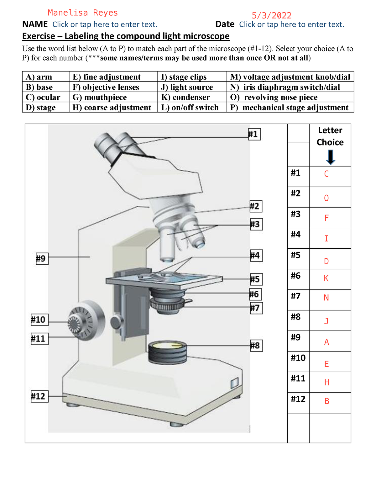
Solved Microscope parts/labeling 9 Label the image of a ...
Microscope Labeled Pictures, Images and Stock Photos Browse 49 microscope labeled stock photos and images available, or start a new search to explore more stock photos and images. Newest results Fluorescent Imaging immunofluorescence of cancer cells growing... Microscope diagram vector illustration. Labeled zoom instrument... Microscope diagram vector illustration.
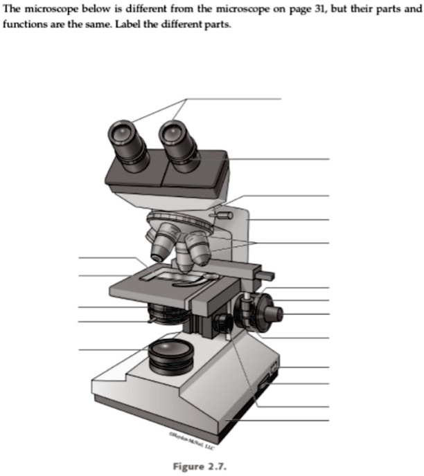
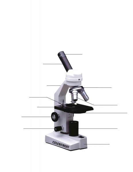
the microscope below i different from the micoscope on page 31 but their parts and functions are the sme label the different parts figure 27 93814
Microscope slide with label royalty-free images - Shutterstock Microscope slide with label royalty-free images 182 microscope slide with label stock photos, vectors, and illustrations are available royalty-free. See microscope slide with label stock video clips Image type Orientation Color People Artists Sort by Popular Healthcare and Medical Science Biology microscope slide medicine microscope pathology

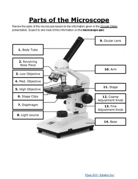
Parts of a microscope with functions and labeled diagram
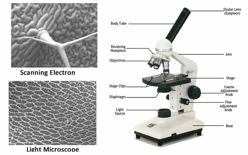
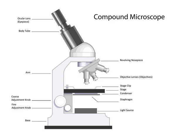
Compound Microscope Parts - Labeled Diagram and their Functions The eyepiece (or ocular lens) is the lens part at the top of a microscope that the viewer looks through. The standard eyepiece has a magnification of 10x. You may exchange with an optional eyepiece ranging from 5x - 30x. [In this figure] The structure inside an eyepiece. The current design of the eyepiece is no longer a single convex lens.

Microscopes for Sale: Compound, Digital & Stereo | NY ...
473,906 Microscope Images, Stock Photos & Vectors | Shutterstock Find Microscope stock images in HD and millions of other royalty-free stock photos, illustrations and vectors in the Shutterstock collection. Thousands of new, high-quality pictures added every day.

Dissecting Stereo Microscope Parts and Functions
Learning to segment microscopy images with lazy labels The need for labour intensive pixel-wise annotation is a major limitation of many fully supervised learning methods for segmenting bioimages that can contain numerous object instances with thin separations. In this paper, we introduce a deep convolutional neural network for microscopy image segmentation. Annotation issues are circumvented by letting the network being trainable on coarse labels ...

National Ecoline D-ELDB Binocular Digital Microscope
Learning to segment microscopy images with lazy labels In this paper, we introduce a deep convolutional neural network for microscopy image segmentation. Annotation issues are circumvented by letting the network being trainable on coarse labels combined with only a very small number of images with pixel-wise annotations. We call this new labelling strategy `lazy' labels.
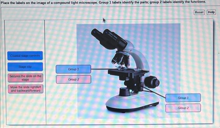
Solved Place the labels on the image of a compound light ...
› Magnifying-Magnifier-HeadbandAmazon.com: Magnifying Glasses 8X 15X 23X Magnifier LED ... About this item . Double eye magnifying glasses magnifier loupe, with 2pcs adjustable LEDs to help it work in low-light conditions ; Left right double eye patches magnifierloupe with adjustable LED to help work in low-light conditions.Set of 2 magnifying glasses mounted on a one-size-fits-all eyeglass frame for easy hands-free operation.
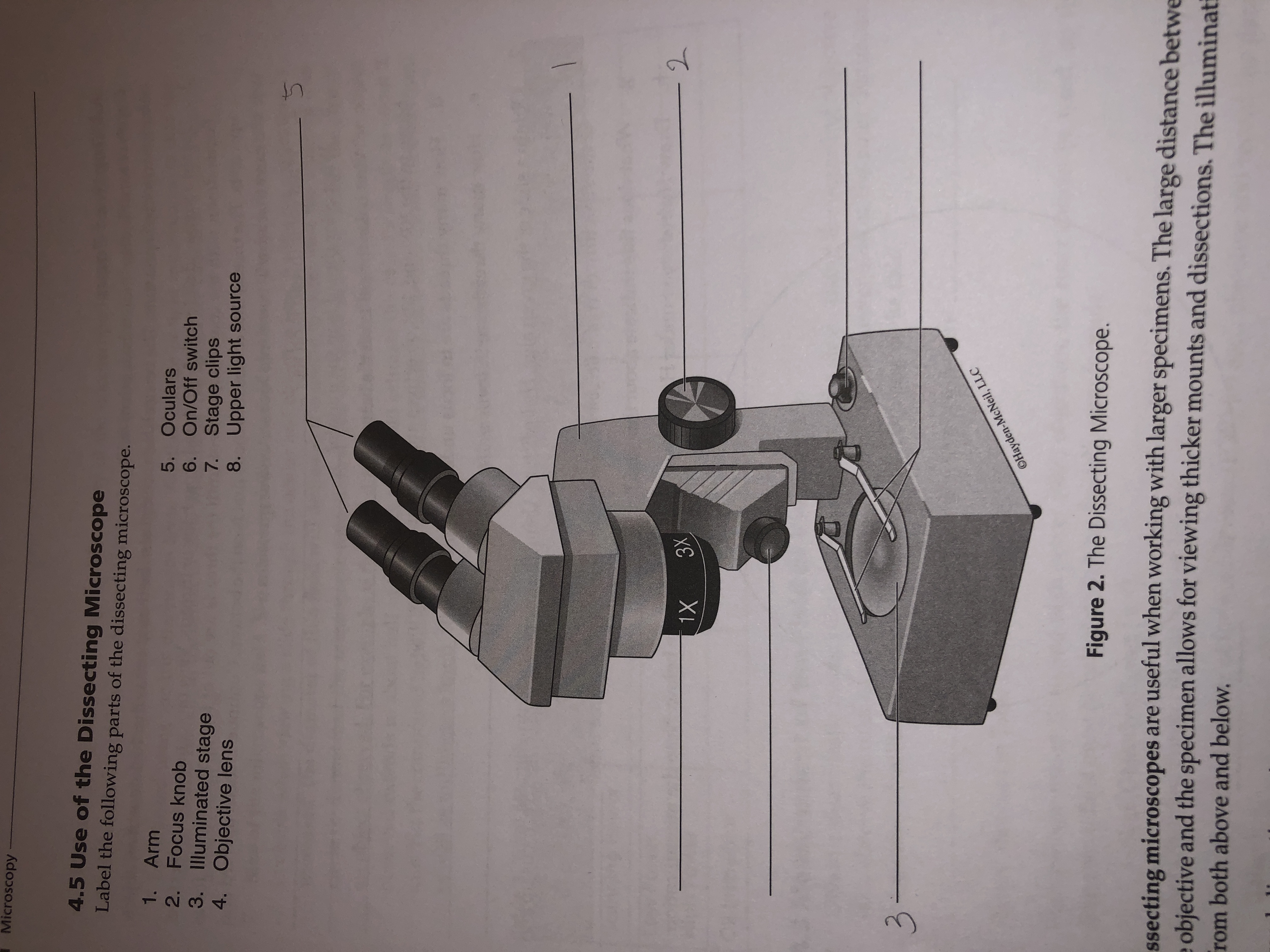
Answered: Microscopy 4.5 Use of the Dissecting… | bartleby
Microscope Parts and Functions First, the purpose of a microscope is to magnify a small object or to magnify the fine details of a larger object in order to examine minute specimens that cannot be seen by the naked eye. Here are the important compound microscope parts... Eyepiece: The lens the viewer looks through to see the specimen.

A Study of the Microscope and its Functions With a Labeled ...
Microscope With Labels Clip Art Image - ClipSafari Microscope With Labels. by: johnny_automatic. a side view drawing of a microscope with parts labeled. From "The Brain in Space" produced by NASA and stated as PD. This is a completely free image Microscope With Labels that you can download, post, and use for any purpose. Tags: equipment, instrument, lab, laboratory, microscope, nasa
microscope | The Biology Corner
Microscope Images at Various Magnifications | Microscope World Resources The images of Paulownia wood, hair, and frog's blood were captured with a high power compound microscope using a Nikon camera adapter. The compound microscope typically has three or four magnifications - 40x, 100x, 400x, and sometimes 1000x. At 40x magnification you will be able to see 5mm. At 100x magnification you will be able to see 2mm.
Free Microscope Drawing, Download Free Microscope Drawing png ...
Fluorescence Microscopy - Explanation and Labelled Images A fluorescence microscope is used to study organic and inorganic samples. Fluorescence microscopy uses fluorescence and phosphorescence to examine the structural organization, spatial distribution of samples. It is particularly used to study samples that are complex and cannot be examined under conventional transmitted-light microscope.
Microscope Slide Labels
Parts of a Simple Microscope - Labeled (with diagrams) A simple microscope is a very first type of microscope ever created. It consists of simple parts and performs simple functions. Although there are now many advanced microscope types, some applications may still demand the use of a simple microscope. In this article, we are going to discuss the parts and functions of a simple microscope.
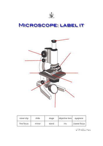
Microscope: label it | Teaching Resources
Labeling the Parts of the Microscope | Microscope World Resources Labeling the Parts of the Microscope This activity has been designed for use in homes and schools. Each microscope layout (both blank and the version with answers) are available as PDF downloads. You can view a more in-depth review of each part of the microscope here. Download the Label the Parts of the Microscope PDF printable version here.

Microscope With Labels clip art | Microscope parts ...
› seterra › en-anThe Heart - Science Quiz - GeoGuessr The Heart - Science Quiz: Day after day, your heart beats about 100,000 times, pumping 2,000 gallons of blood through 60,000 miles of blood vessels. If one of your organs is working that hard, it makes sense to learn about how it functions! This science quiz game will help you identify the parts of the human heart with ease. Blood comes in through veins and exists via arteries—to control the ...
Microscope with labels picture
Labeling the Parts of the Microscope | Microscope activity, Science ... Optical Lens. Microscopic. Compounds. Focal Length. Magnifier. Aperture. Chromatic Aberration. Parts of a Compound Microscope Each part of the compound microscope serves its own unique function, with each being important to the function of the scope as a whole. The individual parts of a compound microscope can vary heavily depending on the ...
Lab - Microscope: MAH-Summer 2019-Anatomy and Physiology I
Microscope Drawing Easy with Label - YouTube In this video I go over a microscope drawing that is easy with label. There is a blank copy at the end of the video to review on your own. A great way to s...

How to Label a Binocular Microscope
› WAI › EMIndex of Dr.Jastrow's electron microscopic atlas Table D leads to images of electron microscopes or protocols for tissue preparation. Table E leads to the overview pages with the images of this atlas which are used in the histology course of the University of Mainz, Germany. From table F you can call up the Vocabulary of microscopic anatomy which explains some terms in German and Englisch.

Fluorescence microscope - Wikipedia
Polarizing Microscope Image Gallery | Science Lab - Leica Microsystems Polarized light microscopy (also known as polarizing microscopy) is an important method used in different fields, including research and quality assurance. It goes beyond just producing images at high magnification and resolution, something typically done with microscopes using ordinary optics. By examining the form, structure, color, birefringence, and further optical properties, additional ...
Print Map Quiz: Labeling the Microscope ()
Compound Microscope Parts, Functions, and Labeled Diagram Compound Microscope Definitions for Labels. Eyepiece (ocular lens) with or without Pointer: The part that is looked through at the top of the compound microscope. Eyepieces typically have a magnification between 5x & 30x. Monocular or Binocular Head: Structural support that holds & connects the eyepieces to the objective lenses.

Microscope Labeling #1 Diagram | Quizlet
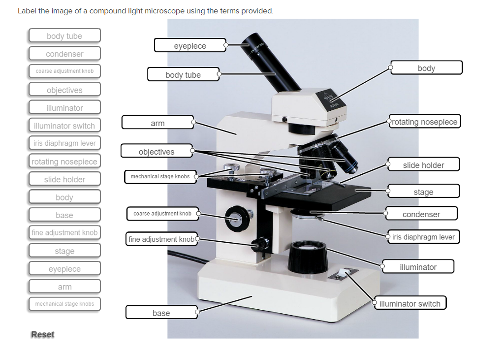
Solved Label the image of a compound light microscope using ...

Labeling the Parts of the Microscope | Microscope activity ...

Microscope Labeling Practice Quiz
Parts of the Microscope Labeling Activity!

Label The Microscope Diagram - Robot PNG Image | Transparent ...
Microscope slide Vector Art Stock Images | Depositphotos
Label microscope - Teaching resources
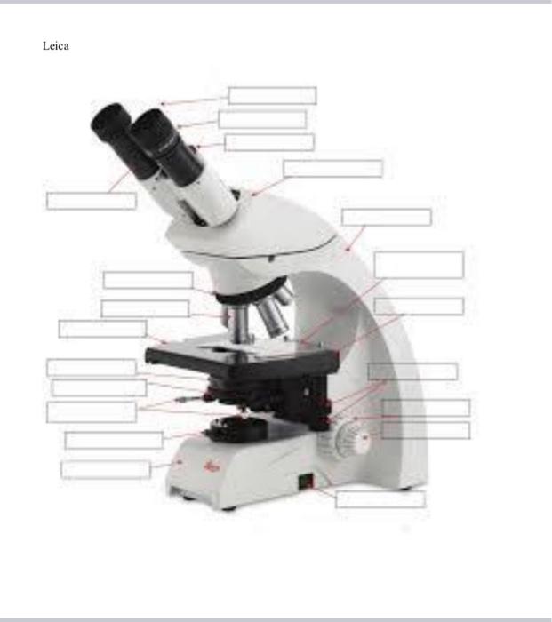
Solved Nikon Parts of the compound microscope Write the ...

Simple Microscope - Diagram (Parts labelled), Principle ...

microscope drawing with label - Clip Art Library

Label the microscope — Science Learning Hub
Color the Microscope Parts
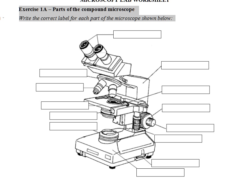
SOLVED: Exercise 1A _ Parts ofthe compound microscope Write ...
BIO 101 parts of the microscope to label - NAME Click or tap ...

Parts of Stereo Microscope (Dissecting microscope) – labeled ...

This is a common compound microscope. Label its parts from A ...

Parts of a Microscope Labeling Activity
Post a Comment for "39 microscope images with labels"